Paola Stephania/Specimen adoption 1: ''Haematococcus Pluvialis''.: Difference between revisions
No edit summary |
No edit summary |
||
| (One intermediate revision by the same user not shown) | |||
| Line 46: | Line 46: | ||
* Day 2: An observation under the microscope is made in order to see the specimen. | * Day 2: An observation under the microscope is made in order to see the specimen. | ||
[[File:hapl0c.jpg|400px]] | [[File:hapl0c.jpg|400px]] | ||
| Line 73: | Line 70: | ||
[[File:hapl2.jpg|400px]] | [[File:hapl2.jpg|400px]] | ||
[[File:hapl0b.jpg|400px]] | |||
[[File:hapl0.jpg|400px]] | [[File:hapl0.jpg|400px]] | ||
| Line 98: | Line 97: | ||
''In the last sample, the concentration of haematococcus can't be seen by the eye, but under the microscope the specimen can be seen as shown in the pictures, also some of the specimens are starting to acquire a green border.'''' | ''In the last sample, the concentration of haematococcus can't be seen by the eye, but under the microscope the specimen can be seen as shown in the pictures, also some of the specimens are starting to acquire a green border.'''' | ||
Conclusions | '''Conclusions''' | ||
1) The environmental conditions for the Haematococcus-pluvialis are not ideal, in order for it to get out of it hibernation state. | |||
My first hypothesis is that it doesn't have the ideal conditions in terms of light and temperature, and my second hypothesis is that it doesn't have the ideal medium ''(a vinasse medium wasn't used for the growth of the specimen, instead a similar medium was made)''. | |||
Latest revision as of 00:04, 4 December 2018
CULTIVATION OF Haematococcus Pluvialis.
- Known by its red color, it's an alga that in stress conditions produces a high content of a strong antioxidant known as astaxanthin.
* SCIENTIFIC CLASIFICATION Division: Chlorophyta Class: Chlorophyceae Order: Chlamydomonadales Family: Haematococcaceae Genus: Haematococcus Species: H. pluvialis Binomial name Haematococcus pluvialis
https://en.wikipedia.org/wiki/Haematococcus_pluvialis
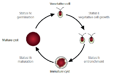
Sequence of growth states of the Haematococcus: https://comunicarciencia.bsm.upf.edu/?tag=haematococcus-pluvialis
- MEDIUM
- 100ml of water
- 3% of vinasse
- 0.7% of NaCl.
The pH is adjusted to 7.0. A 0.4 g/L quantity of inoculum can be used for the initial culture (cells in vegetative growth). The culture must be performed with 0.5 vvm air at 25°C, and until 15 days of culture.
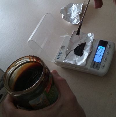
Note: in the laboratory was no Vinasse, so another bioproduct of the sugar was used in order to prepare the medium Malz extract. (brennwert: 1.216kJ, fett 0.2g, kohlenhydrate 65g, Zucker 45g, Eiweiss 5g, Salz0.02g.)
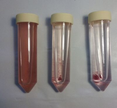
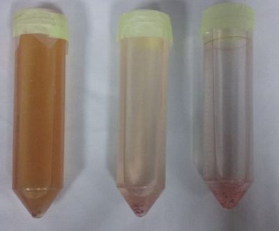
- Day 1: Three samples were made all with the same concentration of salinity but with different percentage of vinasse, and with the same PH 7. Preparation and from left to right; original medium, 10% of original medium diluted in 100ml of water, 1% of original sample diluted in 100ml of water.
The three samples contain:
1 Sample 3,00g malz extract, 100ml water & 0,7g NACl. 2 Sample 0,30g malz extract, 100ml water & 0,7g NACl. 3 Sample 0,03g malz extract, 100ml water & 0,7g NACl.
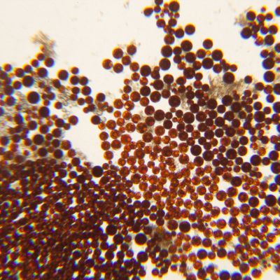
- Day 2: An observation under the microscope is made in order to see the specimen.
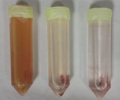
- Day 4: After 4 days of growth the specimens haven't acquired an orange color (this color was expected if the conditions of the environment were ideal for the specimen.
- Day 10: The specimen seems to be starting to attach to the bottom of the tube.
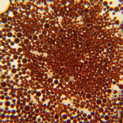
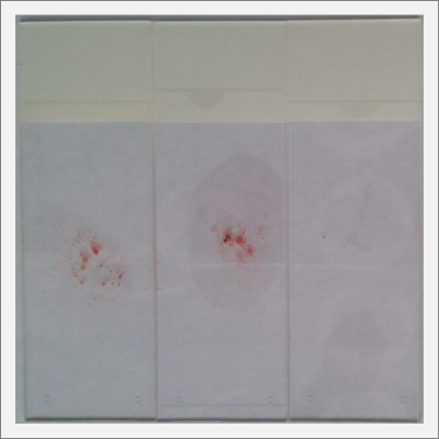
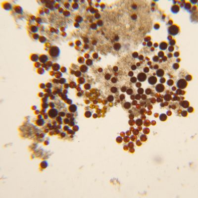
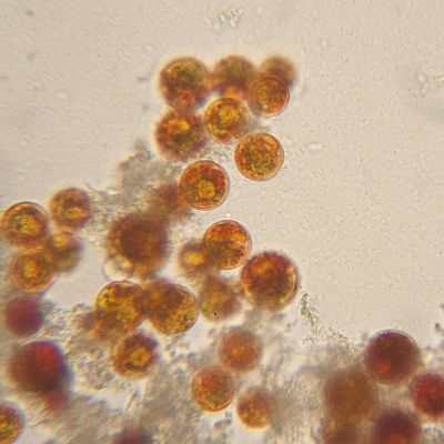
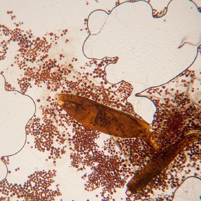
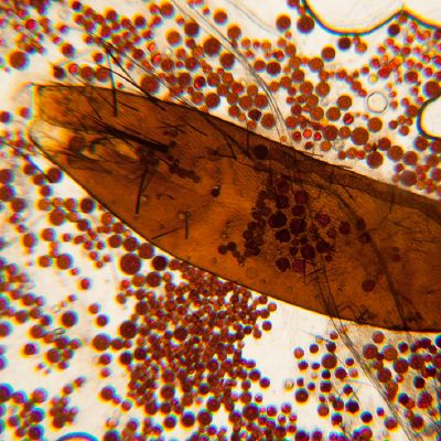
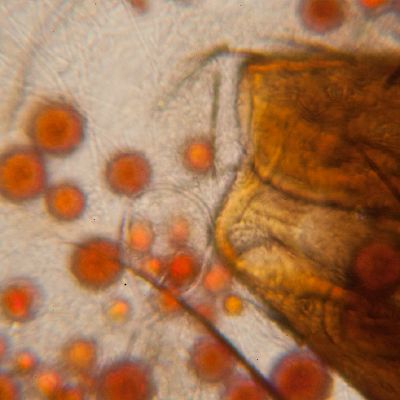
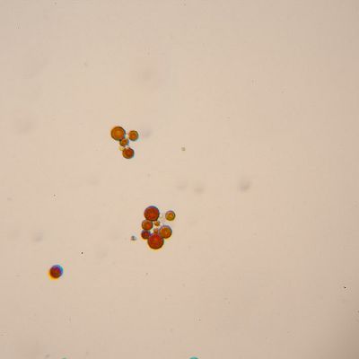
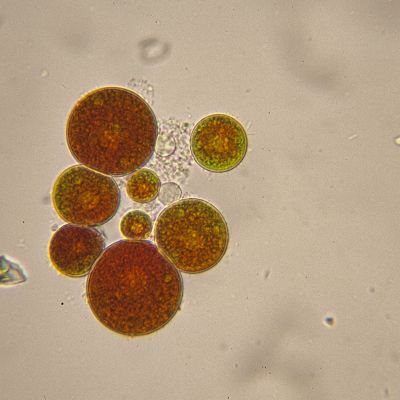
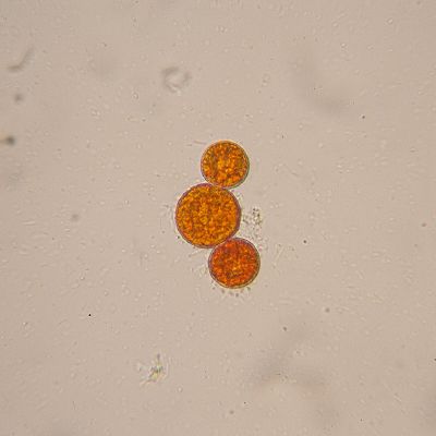
- Day 15: I made an observation of the three samples under the microscope, here are the results:
From left to right more concentration of vinasse and less according to the made samples.
- 1 Sample 3,00g malz extract, 100ml water & 0,7g NACl.
The sample if full with specimens, that can be seen with the eye like a red spot. Under the microscope.
- 2 Sample 0,30g malz extract, 100ml water & 0,7g NACl.
In the sample, I found another specimen, that is probably feating from the Haematococcus pluvialis
- 3 Sample 0,03g malz extract, 100ml water & 0,7g NACl.
In the last sample, the concentration of haematococcus can't be seen by the eye, but under the microscope the specimen can be seen as shown in the pictures, also some of the specimens are starting to acquire a green border.''
Conclusions
1) The environmental conditions for the Haematococcus-pluvialis are not ideal, in order for it to get out of it hibernation state. My first hypothesis is that it doesn't have the ideal conditions in terms of light and temperature, and my second hypothesis is that it doesn't have the ideal medium (a vinasse medium wasn't used for the growth of the specimen, instead a similar medium was made).
