m (→Copper Model) |
|||
| Line 56: | Line 56: | ||
</gallery> | </gallery> | ||
The conductor board material can be easily sawn with a fret saw. Any sharp edges can be removed with rasps and sanding paper. The parts are assembled by soldering them together, also a nut is soldered on the inside of one of the wedges. The threaded bold is fixated to the box with | The conductor board material can be easily sawn with a fret saw. Any sharp edges can be removed with rasps and sanding paper. The parts are assembled by soldering them together, also a nut is soldered on the inside of one of the wedges. The threaded bold is fixated to the box with small clamps to keep it steady but turnable at the same time. It reaches through the outer wall of the box into the nut within the wedge and thereby provides the mechanism for the wedge movement. | ||
== Microscopic Images == | == Microscopic Images == | ||
Revision as of 12:14, 28 March 2013
Miroscopy Stage Construction
Approach with 2 wedges. The aim was to find a simple mechanism that allows for high precision. If you tighten the screw the lower wedge moves left and pushes up the upper wedge with the camera on top of it. If you loosen the screw the camera moves down. Because of its simplicity it would be less susceptible to errors.
3D Model
You can download the model here: Media:ModelWedges.zip. You need to have Google SketchUp installed to open the model.
Preparing the Camera
First remove the lens of the camera. Use a screwdriver to unscrew the outer parts. Then use a hand saw or a pair of nippers to crack open the case around the cable. Carefully remove the plastic without damaging the cable. As a result you get the PCB with the cable and the LEDs. For now leave the black plastic ring on the PCB as a protection for the sensor.
The second step is to remove the LEDs. Unsolder them carefully and clean the solder holes with a desoldering pump.
Cardboard Model
Copper Model
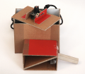
After checking and improving the functionality of the design with the cardboard construction the final model can be built. Here the base material for conductor boards is used. It is made out of epoxy with a copper surface. This makes the material easy to work on. The smooth surface of the copper allows for a smooth movement of the wedges. The design was expanded by two small springs that constantly pull down the camera platform, preventing the platform to be pushed up by the firm cables of the camera. What you need is:
- conductor board material
- threaded bolt
- plastic knob
- soldering rod
- solder
- two small extention springs
- small metall clips to keep the threaded bolt in place
- ribbon cable
- tiny screws
- plastic spacers to fixate the camera in the right place
- fretsaw
- rasp
- sanding paper
- drill
The conductor board material can be easily sawn with a fret saw. Any sharp edges can be removed with rasps and sanding paper. The parts are assembled by soldering them together, also a nut is soldered on the inside of one of the wedges. The threaded bold is fixated to the box with small clamps to keep it steady but turnable at the same time. It reaches through the outer wall of the box into the nut within the wedge and thereby provides the mechanism for the wedge movement.
Microscopic Images
Calculating Camera Resolution
An iPad with 52ppcm (pixel per cm) was used to calculate the camera resolution. This means that one pixel is 10/52 mm wide, that is roughly 0,2 mm. These 0,2 mm equal 300 pixels of the camera image. Therefore one pixel of our camera shows 0,00067 mm (0,2mm/300) of the specimen. Knowing that the image is 640 pixels wide and 480 pixels high, the image size adds up to 0,43 mm in width (0,00067*640) and 0,32 mm in height (0,00067*480).
Artemia Salina
With the objective of finding interesting material to examine Artemia Salina were breeded. Artemia Salina is a very old species of brine shrimp. Breeding sets can be found in the pet shop and have been a gimmick in childrens science magazines on a regular basis. Unfortunately even the newly hatched shrimps called nauplii are to big to be examined thoroughly with the camera.
<videoflash type=vimeo>54231452|400|300</videoflash>
Hay infusion
Another possibility to create specimen to look at with a microscope is the hay infusion. Collect a handfull of hay and dry leaves and about 500 ml of pond water. Put the hay in a glass bowl and pour the water on it. Keep your hay infusion in a bright place at room temperature. After a few days you can find protozoa like Paramecium, Amoebia or Euglena. The hay infusion results in different protozoa depending on the material that is used. Possible materials are hay, dry leaves or salad that wasn't treated with pesticides.
After 5 days at room temperature a lot of bacteria and also quite a lot of protozoa have developed. <videoflash type=vimeo>54410341|400|300</videoflash> <videoflash type=vimeo>54232064|400|300</videoflash>
Clotting Blood
This is a fast motion video of clotting blood. The video's speed was increased by about 700%. The real time clotting took almost 5 minutes.
<videoflash type=vimeo>54409738|400|300</videoflash>
Guess what
<videoflash type=vimeo>54253797|400|300</videoflash>
Artistic approach
Image to Sound
The aim is to take different parameters of the microscopic picture and use them to generate sound. To achieve this the visual programming language Pure Data and the software Mainstage are used. The chosen parameters are brightness, movement and the histogram of the picture.
So far brightness and the histogram are transformed into sound. The videos show the histogram and the corresponding picture.
<videoflash type=vimeo>56874602|520|290</videoflash> <videoflash type=vimeo>56874349|520|290</videoflash> <videoflash type=vimeo>56874082|520|290</videoflash> <videoflash type=vimeo>56874080|520|290</videoflash>


