| Line 11: | Line 11: | ||
===''First culture medium''=== | ===''First culture medium''=== | ||
As described obove the filled petri dishes are no beeing observed for the following days. | As described obove the filled petri dishes are no beeing observed for the following days. | ||
====Gallery==== | ====''Gallery''==== | ||
<gallery | <gallery mode="nolines"> | ||
File:petrischale mit Nährmedium.jpg|Day 1 | File:petrischale mit Nährmedium.jpg|''Day 1'' | ||
File:Tag 3_Nährmedium.JPG|Day 3 | File:Tag 3_Nährmedium.JPG|''Day 3'' | ||
File:Tag 6_Nährmedium.JPG|Day 6 | File:Tag 6_Nährmedium.JPG|''Day 6'' | ||
File:Tag 7_Nährmedium.jpg|Day 7 | File:Tag 7_Nährmedium.jpg|''Day 7'' | ||
</gallery> | </gallery> | ||
Revision as of 12:36, 22 October 2018
1st class: 16. Oktober 2018
Summary
Preparing the first testing Agar-Plates for bacterial growth in a petri dish:
- petri dishes where filled with 15 ml of an LBP Medium with Agar Agar
- cooling down
- inoculateing with different surface smear
- In my case: human nose (inside), cat nose (outside) and fur, fridge glass surface and 1 cent coin
- left with room temperature for the following days
First culture medium
As described obove the filled petri dishes are no beeing observed for the following days.
Gallery
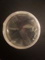
Day 1
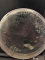
Day 3
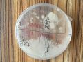
Day 6
Day 7


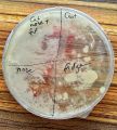
breeding day 7 - closer look
Somehow I got the impression that the petri dishes weren't sterile as after 7 days almost the full dish is covered with a white film. You can only guess by slightly different colors where I initially inoculated the dish. Something seems to grow over the originally inoculated sections. See below some close ups. In the section of the example I took from my nose, a small yellow dot appears. I think this is what was intended to be growing. The rest just looks almost the same.
Gallery

Cat fur and outside nose

1 Euro cent


Day 7 - Full dish

Human nose inside

glass surface fridge


DIY Microscope Test
What was done?
- extracted converging lens from a laser pointer
- attached to iPhone SE back camera

Chestetree seed

Sunflower seed

Sunflower and chestetree seed in comparison

Chestetree seed magnified

Sunflower seed magnified


- adding some filters to increase contrast und depth

Artsy chestetree seed

Artsy sunflower seed

