HUMAN DNA ANALYSIS
I grew up with the fantasy that I must have something exotic deep inside me. People frequently asked me about my relatives, and I started to believe that I somehow belong to another soil than my motherland. Having the chance to work in a DIY Biolab, I decided to pursue my intersect feelings and I attended to analyze my DNA sequences, with the secret hope of discovering my own ethnicity extent and finding any connection with Asian pattern genome.
After making myself familiar with this new terminology, I have been gathering different protocols which would allow me to identify a certain percentage of Asian patterns in my DNA sequences. But unfortunately, I found out that it was more complex than I first thought, for multiple reasons. The first is the fact that those protocols required a more specific equipment (than PCR machine), but then and above all, an access to a DNA database is indispensable. Indeed, the overall principle of each method is about comparing DNA samples with an already defined categorized population guide. In addition, the amount of genetic variations among different population group is 10%-20% only, and within a population component of genetic variation of 93% to 95% has been established.
Thus, categorizing a group of population is already a long, complex and tricky method, but then, finding fragments of different ethnicity patterns within a single DNA sample turns towards a speculative work. Plus, even if I would have found some significant Asian genetic patterns in my blood, those very patterns would have been compared with actual Asian population, and not with an ancestral one (which could differ)
PRACTICING PCR ANALYSIS
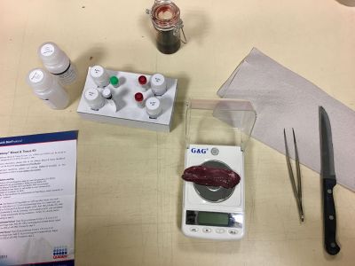
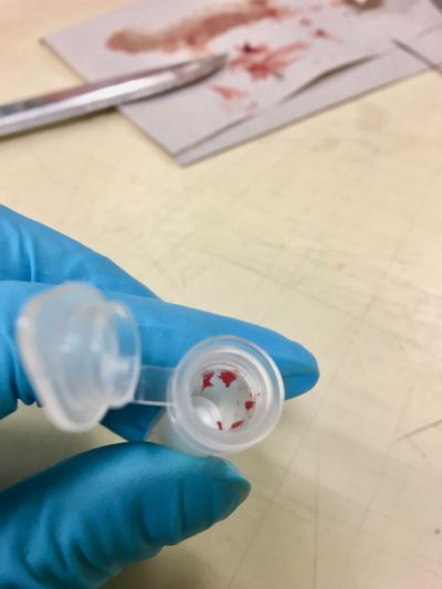
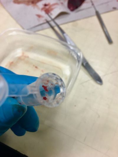
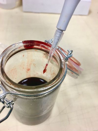
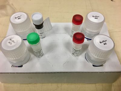
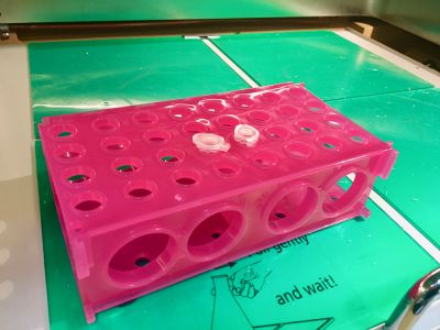
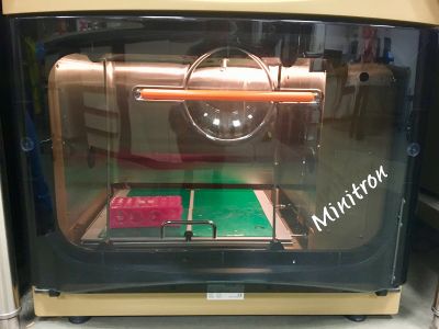
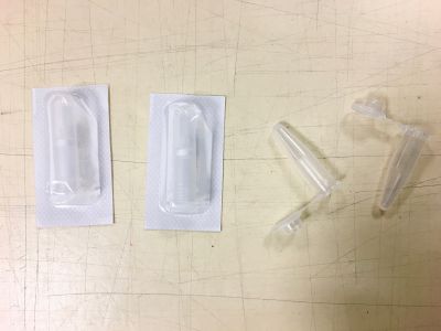
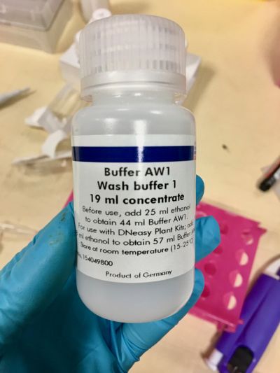
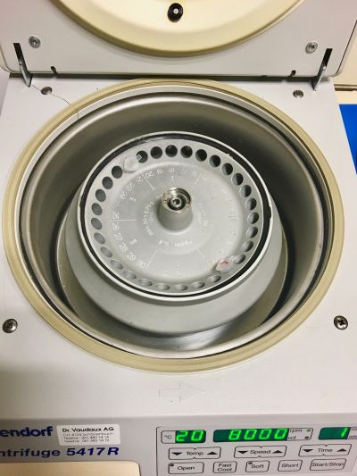
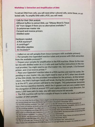
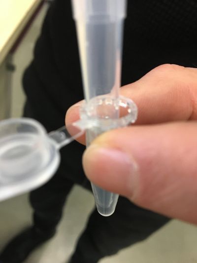
Step 1. Extracting DNA from blood and from pork liver meat
Step 2. STR-analysis with pork meat primer
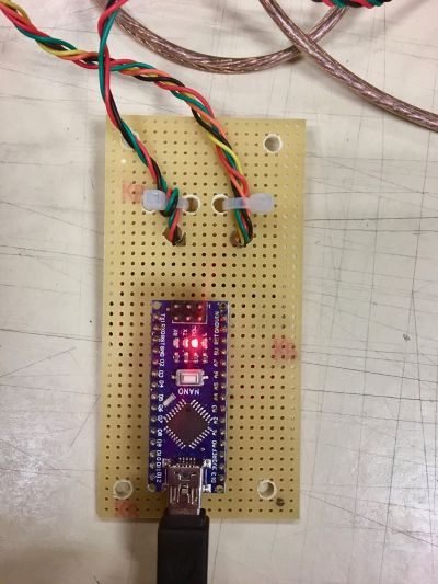
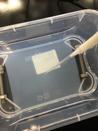
Step 3. Electrophoresis
#First attempt
=> I got no results: either the ladder nor the DNA samples were "growing" as they should (multiple lines). So I decided to test the ladder only to see if there is a problem with the samples themselves or with the preparation (agarose + buffer TAE 50x+ distilled water) => #Second attempt
#Second attempt
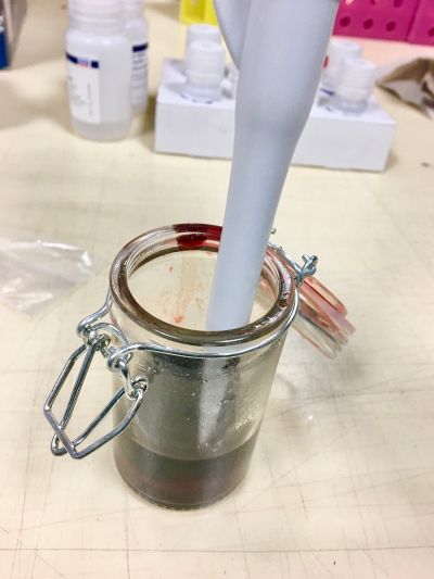
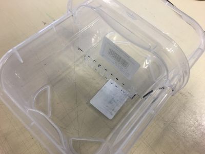
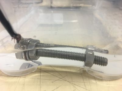
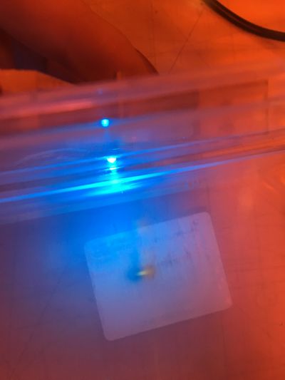
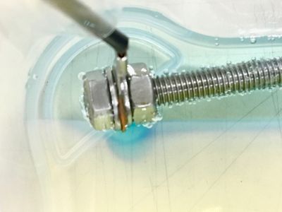
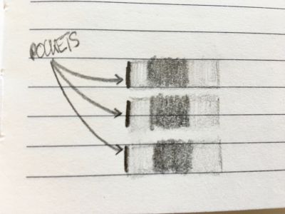
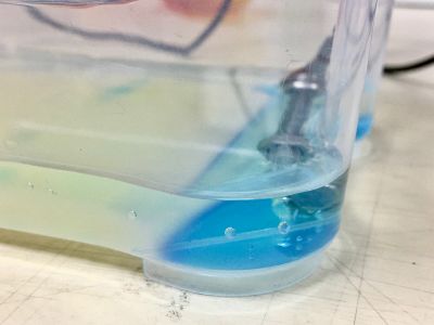
=> Same problem: the ladder was growing as one bloc only (see the little drawing). Even during a 2.2 attempt with new SERVA Stain G (which I thought it could be the problem since the previous tube was quite old). Then, I noticed that the positive side was turning in blue color and some part of the agarose gel was melting down. So I decided to test if this blue color is coming from oxidation of the electrodes themselves, or if the ladder was "swimming" on the extra amount of solution (buffer TAE 50x + distilled water) above the agarose to reach the positive side => #Third attempt
#Third attempt
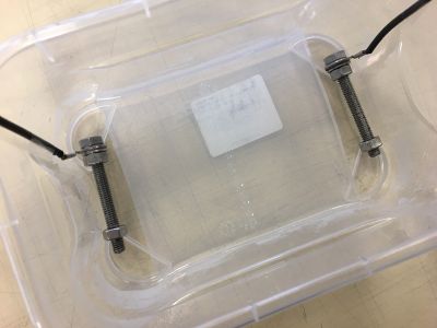
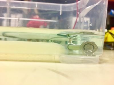
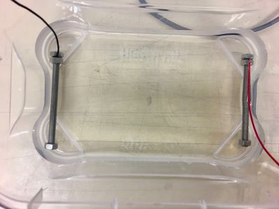
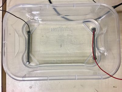
=> Still turning blue. Even with smaller electrodes that are completely covered by the solution (buffer TAE 50x + distilled water) So I decided to test if this oxidation is coming from the buffer or if it is simply a natural process => #Fourth attempt
#Fourth attempt
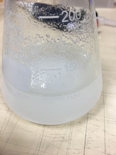
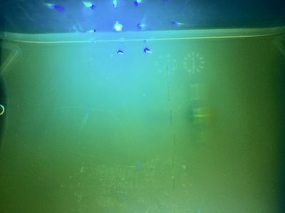
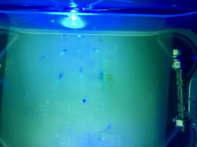
=> First picture: distilled water only. Second picture: distilled water + buffer TAE 50x. No changes at all. So it might come from the SERVA Stain G (which is yellow-ish) or it is actually not a problem and does not affect the electrophoresis itself.


